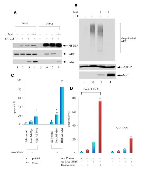
Figure 2. Distinct Effects of Different c-Myc Expression Levels on ULF and ARF.
(A) Higher levels of c-Myc, but not lower, block the interaction between ULF and ARF. In order to better monitor the binding affinity, the cells were pretreated with proteasome inhibitors. Western blot analysis of crude cell extracts and M2-immunoprecipitates from human 293 cells transfected with expression vectors of ARF, FH-ULF (FH: Flag and HA tagged) and low to high amounts of c-Myc (1:5 ratio) in the presence of a proteasome inhibitor epoxomycin by anti-HA for ULF, anti-ARF and Myc antibodies.
(B) Higher overexpression of c-Myc, but not lower, inhibits ARF ubiquitination mediated by ULF. Human 293 cells were cotransfected with vectors encoding HA-tagged ubiquitin, ARF, ULF and low to high amounts of Myc (1:5 ratio) as indicated. Lysates were immunoprecipitated with anti-ARF antibody (ab-4, Labvision), and proteins were blotted with antibodies to the HA-tag (top) or ARF (middle). Ectopic Myc in crude lysates was analyzed by western blot with 9E10 antibody (low).
(C) Apoptotic cells (Sub-G1) according to DNA contents (PI staining) were counted for the adenoviral MYC infected NHF cells, plus those cells with additional 0.2 mg/ml doxorubicin treatment by FACS. Error bars represent s.d. (n=3).
(D) Apoptotic response mediated by DNA damage in high Myc overexpression NHF-1 cells is ARF-dependent. Apoptotic cells (Sub-G1) according to DNA contents (PI staining) were counted for ARF RNAi treated NHF-1 cells followed by 36 hrs of Ad-Myc virus (MOI 50) infection, and subsequent 24 hrs of 0.2 mg/ml doxorubicin treatment by FACS. Error bars represent s.d. (n=3). See also Figure S2.

