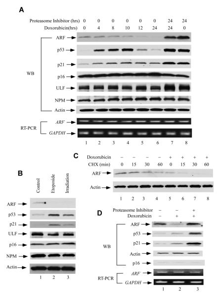
Figure 3. DNA Damage Induces an Ubiquitination-Dependent Human ARF degradation.
(A) Western blot analysis of cell extracts from normal human fibroblast cells (NHF-1) harvested at indicated time points after 0.2 mg/ml doxorubicin and/or 100 nm epoxomycin treatment by anti-ARF, p53, p21, p16, ULF and NPM antibodies. ARF and GAPDH mRNA expression levels by RT-PCR were shown at lower panels.
(B) Western blot analysis of cell extracts from NHF-1cells treated with 20 μm etoposide (lane 2) for 24 hrs, or 5Gy ionizing radiation (irradiation) (lane 3) versus control (lane 1).
(C) Western blot analysis of cell extracts by an anti-ARF antibody from control (lanes 1-4) or NHF-1 cells treated with doxorubicin for 20 hrs (lane 5-8), and followed by cyclohexamide (CHX) treatment (min).
(D) Western blot analysis of cell extracts from normal human IMR90 cells treated with 0.2 mg/ml doxorubicin (lane 2), together with 100 nm epoxomycin (lane 3) or control (lane 1) by anti-ARF, p53, p21 and p16 antibodies. ARF and GAPDH mRNA expression levels by RT-PCR were shown at lower panels. See also Figure S3.

