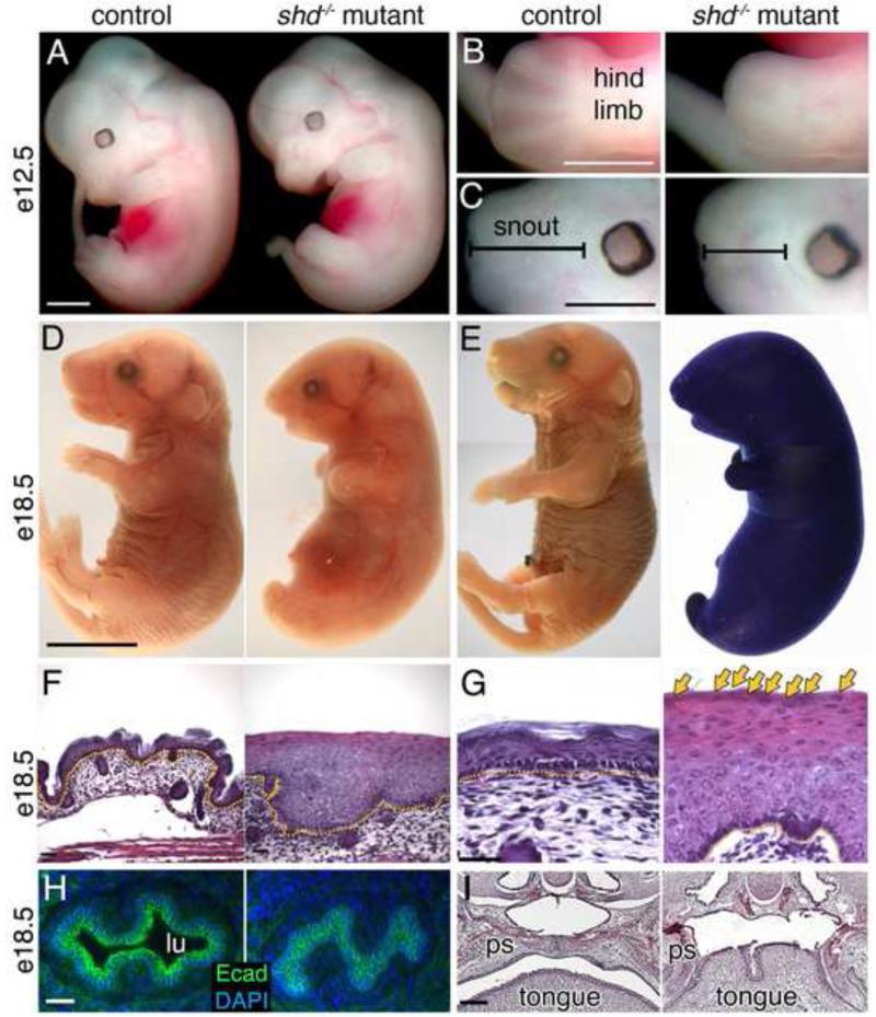
Figure 1. shd mutants display multiple defects linked to keratinocytes.
(A-C) Gross morphological comparison of wild-type and shd mutant embryos at e12.5. shd mutants display shortened and fused limbs (B) and craniofacial malformations, including shortened snout (C). (D) Gross appearance of wild type and shd mutants at e18.5, showing a smooth skin and fusion between hindlimbs and tail. (E) Toluidine Blue exclusion assays of whole embryos show a breakdown in the epidermal barrier in shd homozygotes. (F-G) Hematoxylin and eosin-stained sections of back skin showing a thickened epidermis in shd mutants and retention of nuclei in distal keratinocytes (G, arrows). (H) Sections through the esophagus reveal an absence of a lumen (lu) in shd homozygotes and apparent fusion between the keratinocytes (E-Cadherin+) lining the esophagus. (I) Hematoxylin and eosin-stained sections through heads showed that the palatal shelves (ps) do not fuse along the midline in shd mutants, but instead appear fused to the tongue, resulting in cleft palate. Scale bars: 1mm (A-C) 5mm (D, same for E), 50um (F,G), 20um (H), 100um (I). Yellow dotted lines in F, G mark the dermis (lower) and epidermis (upper) boundary.

