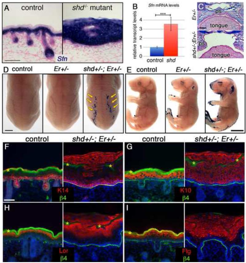
Figure 3. Er-shd double heterozygotes develop cleft palate and epidermal differentiation defects.
(A) in situ hybridization for Sfn (blue) at e18.5 shows expanded expression in shd epidermis. (B) qRT-PCR reveals elevated Sfn transcript levels in shd epidermis. (C) Alcian blue and Fast Red-stained sections through e18.5 heads show that the palatal shelves (ps) elevate and fuse normally in Er heterozygotes but do not fuse along the midline in shd-Er double heterozygotes, resulting in cleft palate. (D,E) Dye exclusion assays show a delay in barrier formation in Er heterozygotes and a more pronounced epidermal barrier defect in shd+/−;Er+/− animals including stripes in caudal back skin (D) and around the eyes, ears, snout, limbs and tail (E). Sections through the barrier-defective dorsal epidermis show that the epidermis is thickened in these regions (F-I) and that basal (F, K14) and spinous (G, K10) markers are abnormally expanded in distal shd+/−;Er+/− keratinocytes. Terminal differentiation markers (E,F) are expressed in shd+/−;Er+/− keratinocytes, but are co-expressed in with K14 and K10. β4 integrin staining marks the border between the epidermis and dermis (F-I). DAPI is shown in blue (F-I). Scale bars represent 50um (A), 250um (C), 2mm (D), 5mm (E), 20um (F-I).

