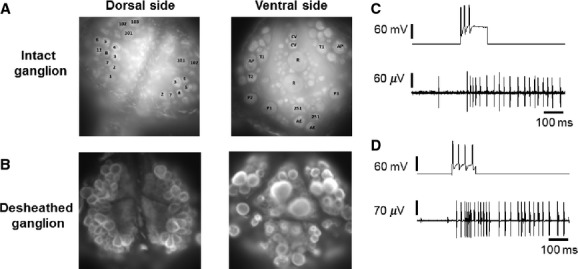
Figure 1.

Preparation. (A) images of a dorsal and a ventral surface of an intact leech ganglion, stained with the voltage-sensitive dye VF2.1.Cl. (B) as in A, but after desheathing the ganglion. (C) Upper trace: intracellular recording from a P cell in an intact ganglion, during which the sensory neuron was depolarized by injecting a pulse of current through the microelectrode. The injected current pulse evoked a train of spikes in the sensory neuron, which elicited a train of spikes recorded extracellularly with a suction electrode from a DP nerve (lower trace). (D) as in C but in a desheathed ganglion; a train of spikes in a P cell evoked a train of spikes in the DP nerve.
