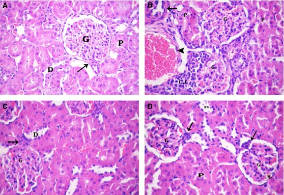
Figure 3.

Renal cortex of: (A) Control group showing renal corpuscle with normal glomerulus (G). The juxtaglomerular apparatus (↑) is seen. Notice normal pattern of proximal convoluted (P) and distal convoluted (D) tubules. (B and C) Salt-loaded group: (B) showing most of the corpuscles (G) with high cellularity and obliterated capsular space. Proximal convoluted tubules (P) show destructed epithelial lining. Notice congested blood vessels (▲), thickened arterioles (↑), and cellular infiltration (C.I.). (C) Showing high cellularity of renal corpuscles (G) and extra-mesangial cells (↑) of juxtaglomerular apparatus. Notice destructed epithelial lining of distal convoluted tubules (D). (D) Omega-3-treated salt-loaded group showing preservation of normal pattern of renal corpuscles (G) and juxtaglomerular apparatus (↑). Both proximal (P) and distal (D) convoluted tubules appear nearly normal (H&E 400 × ).
