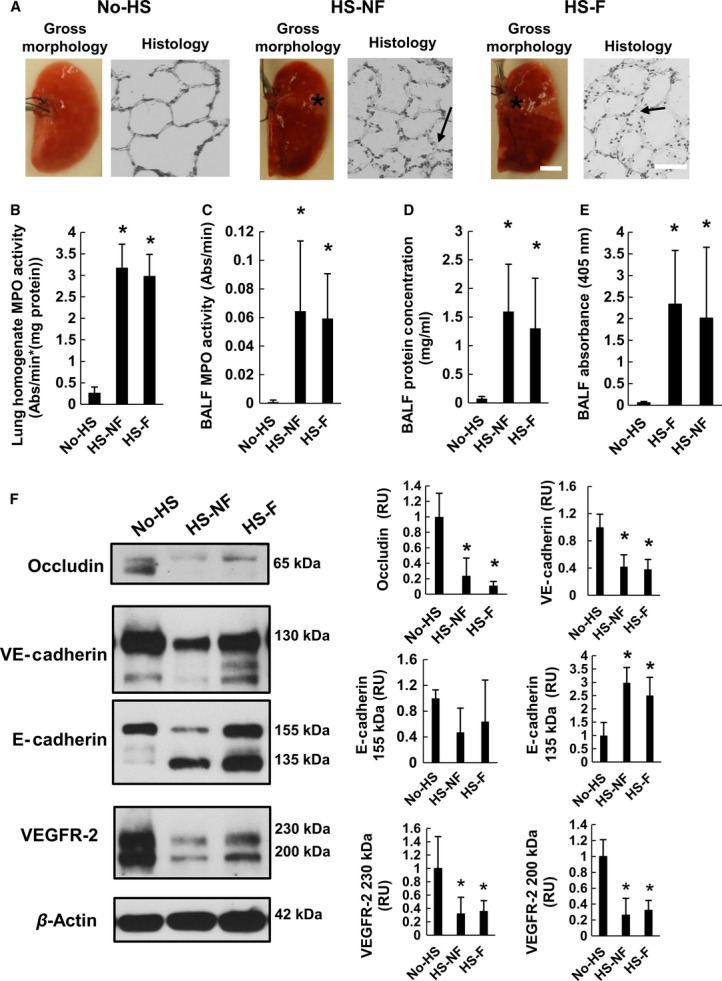
Figure 6.

Markers for lung injury after hemorrhagic shock (HS). (A) Lung damage in form of microhemorrhages (see *) as observed by gross morphology. Histological sections of the most damaged lungs had visible swelling of the alveoli (arrows) in corresponding sample images magnified on the right. No-HS control exhibited no bleeding. (B) Lung MPO activity. (C) BALF protein concentration. (D) BALF MPO activity. (E) BALF absorbance were elevated after shock regardless of whether or not the intestine was flushed. (F) VE-cadherin, E-cadherin, occludin, and VEGFR-2 main isoforms in lung homogenates detected by immunoblot. They decrease following HS. Band densities are standardized to β-actin bands and normalized by No-HS levels. *P < 0.05 by ANOVA followed by Tukey post hoc. Bar graphs show mean ± SD. N = 6 rats/group.
