
Figure 2.
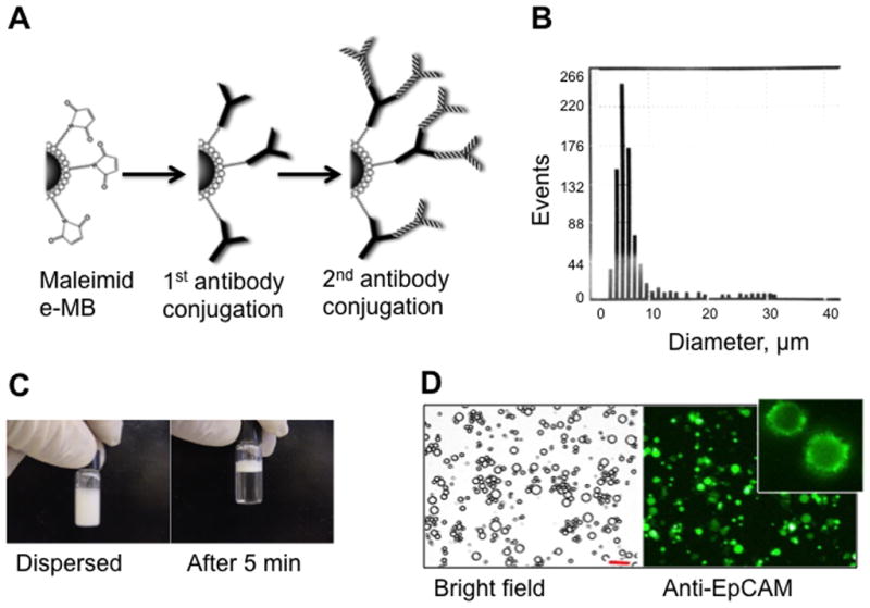
Preparation and characterization of MBs. (A) Maleimide-MBs are coated with anti-Fcspecific antibody and anti-EpCAM antibody in a two-step process. (B) Resulting MBs have a broad size distribution, as measured with Countess™ cell counter software. (C) MBs float soon after redispersion. (D) Microscope images of the resulting MBs after staining of anti-EpCAM IgG with Alexa 488-labeled secondary antibody. Inset shows cropped image of a MB with characteristic “dentate” fluorescence. Size bar 20 μm.