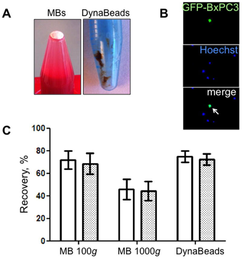
Figure 5.

Isolation of GFP-positive BxPC3 pancreatic carcinoma cells from 1 ml blood with anti-EpCAM MBs and magnetic beads. (A) MBs after centrifugation form white pellet on top of the liquid. DynaBeads form slurry on the side of the tube after washing with external magnet. (B) Tumor cells are distinguished from leukocytes due to GFP/Hoechst label (arrow). (C) Effect of centrifugation speed on the recovery of BxPC3 cells from BSA/PBS (white bars) or blood (grey bars). Isolation efficiency in the MB layer decreased with 1000g force, possibly due to the tumor cell detachment from MBs. At 100g, the isolation efficiency was similar to DynaBeads.
