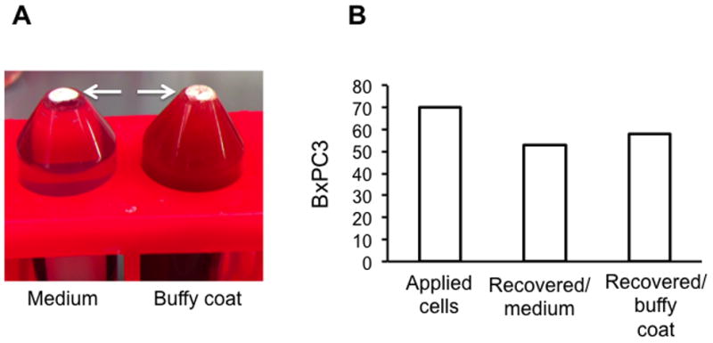
Figure 6.

Isolation of spiked tumor cells from large volume of buffy coat or medium. (A) BxPC3 cells (non-GFP) were spiked into 22 ml medium (left tube) or 22 ml plasma-depleted buffy coats (right tube) and isolated with MBs. Arrows point to the MB layer. (B) Isolation (recovery) efficiency was determined after staining with cytokeratin (CK) antibody and Hoechst and counting CK+/Hoechst+ cells with fluorescent microscope.
