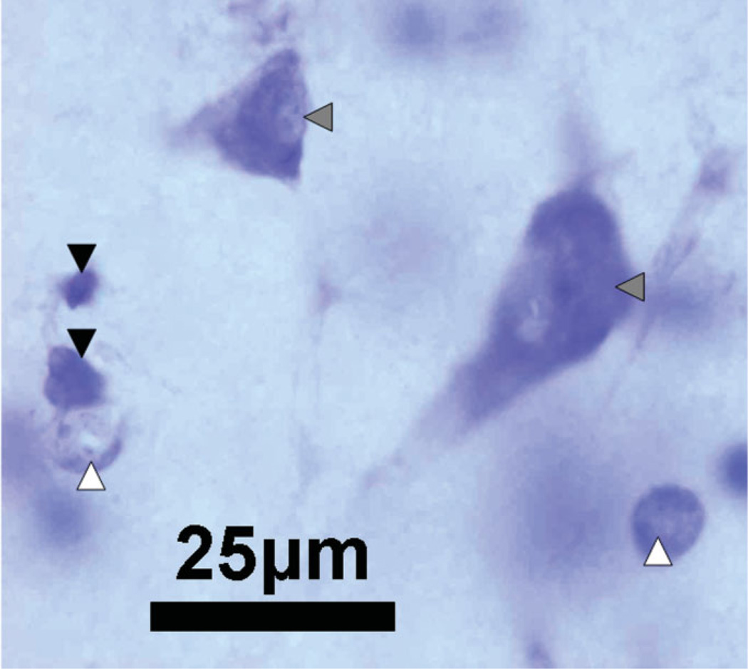
FIGURE 2.
A photomicrograph of the hilus of the dentate gyrus at ×100 magnification, illustrating typical cell types based on morphology and Nissl staining. Neurons, indicated by the solid gray arrowheads, are stained with a large nucleus and a single nucleolus. Astrocytes, indicated by the solid white arrowheads, display pale staining of the nucleus. Oligodendrocytes, indicated by the solid black arrowheads, are identified by dark nuclei; Calibration Bar: 25 µm. [Color figure can be viewed in the online issue, which is available at www.interscience.wiley.com.]

