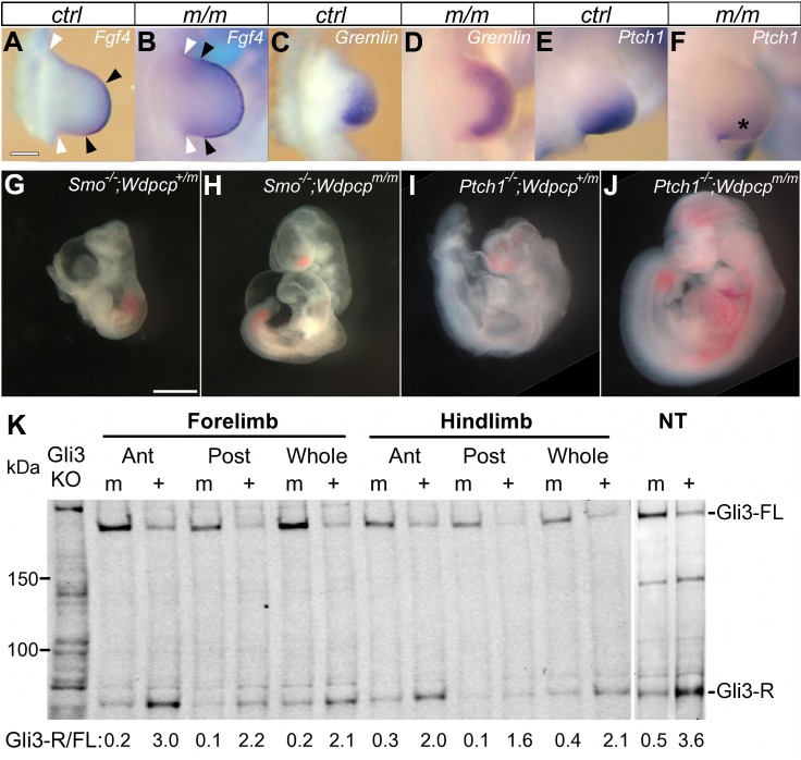
Figure 5. Sonic hedgehog signaling defect in WdpcpCys40 mutant.
(A–F) In-situ hybridization of E10.5 forelimbs shows WdpcpCys40 mutants with expanded expression of Fgf4 in the AER (apical ectodermal ridge) (A, B) and Gremlin in the limb mesenchyme (C, D), but reduced expression of Ptch1 (E, F and asterisk in F). In (A) and (B), black arrowheads indicate the span of the AER, and white arrowheads are the anterior and posterior bases of the limb bud. (G–J) Wdpcp deficiency rescued the severe defect phenotypes of Smo −/− (G) and Ptch1 −/− (I) mutant embryos at E10.5 dpc. The WdpcpCys40/Cys40;Smo −/− (H) and WdpcpCys40/Cys40;Ptch1 −/− (J) double homozygous mutant embryos collected at E10.5 dpc show more robust growth with better axial development and also more normal head and heart development. (K) Western blotting of Gli3 in tissue extracts obtained from the limb and neural tube shows a decrease of Gli3-R/Gli3-FL ratio in WdpcpCys40 mutant embryos. Scale bars, 200 µm in (A), 1 mm in (G). Scales are the same in (A–F) and (G–J).

