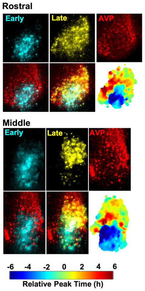
Figure 2. Photoperiodic reorganization of SCN shell and core regions.
Coronal SCN slices collected from LD20:4 mice were imaged for 2 days and then processed for arginine vasopressin (AVP, red)-immunoreactivity. Individual still images of PER2::LUC expression were selected to isolate the early- (blue pseudocolor) and late-peaking (yellow pseudocolor) regions on the first cycle in vitro before superimposition onto AVP-ir images. The results indicate that LD20:4 reorganizes the SCN into shell and core regions cycling in anti-phase, since the AVP-ir neurons that demarcate the SCN shell are in spatial registry with the late-peaking region but not with the early-peaking core-like region. Individual phase maps are indicated for each slice. See also Figure S2.

