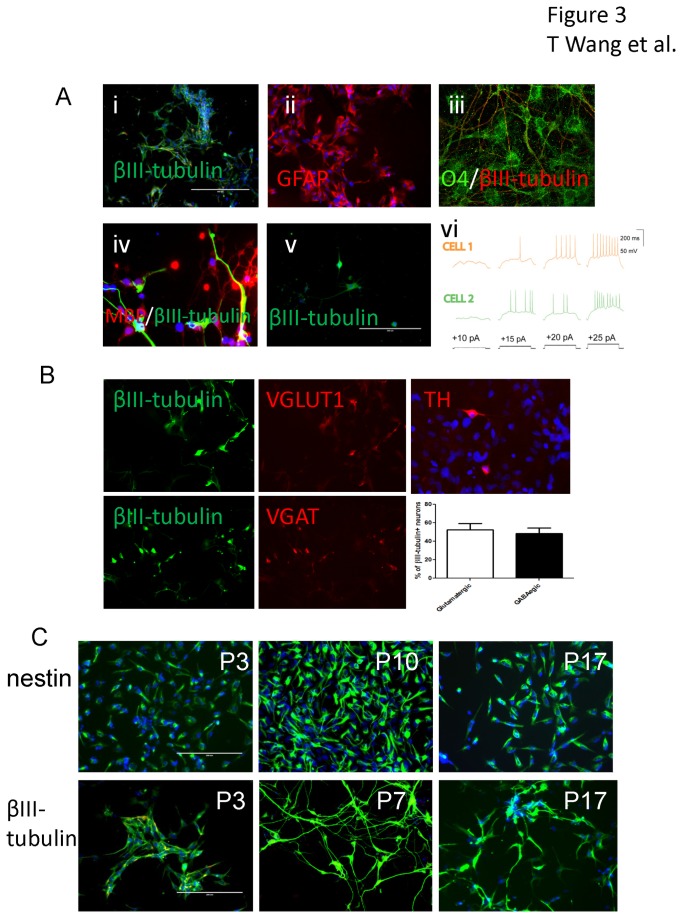
Figure 3. Neurons with action potentials and glial cells differentiated from induced neural stem cells from adult peripheral CD34+ cells.
The induced neural stem cells differentiated into βIII- tubulin positive neurons (green, Ai) in neuronal differentiation or GFAP positive astroglia (red, Aii) in astroglial differentiation medium for 1 week. Oligodendrocyte progenitor differentiation was achieved by incubating the neural stem cells with oligodendrocyte differentiation medium. O4 positive cells (green, Aiii, after 2 weeks) and a few of MBP positive cells (red, Aiv, after four weeks) were detected by immunostaining. Spontaneous neuronal differentiation was also observed in low concentration seeded neural stem cell culture (1000 cells/well in a 24 well plate) after incubation in neural stem cell medium for 1 week (Av). Action potentials were recorded from neurons differentiated for 2 weeks using whole cell patch clamp. Action potentials recorded on two separated cells are shown (Avi). Double staining with either VGLUT1 or VGAT with βIII- tubulin showed most βIII- tubulin positive neurons were either glutamatergic or gabaergic neurons while a few of doparminergic neurons were also detected by TH immunostaining after incubation in neuronal differentiation medium for 2 weeks (B). The induced neural stem cells were passaged for 17 passages and immunostained for nestin as a neural stem cell marker. The cells were also cultured in neural differentiation medium for 2 weeks and immunostained for βIII-tubulin to determine their neuronal differentiation capabilities (C).

