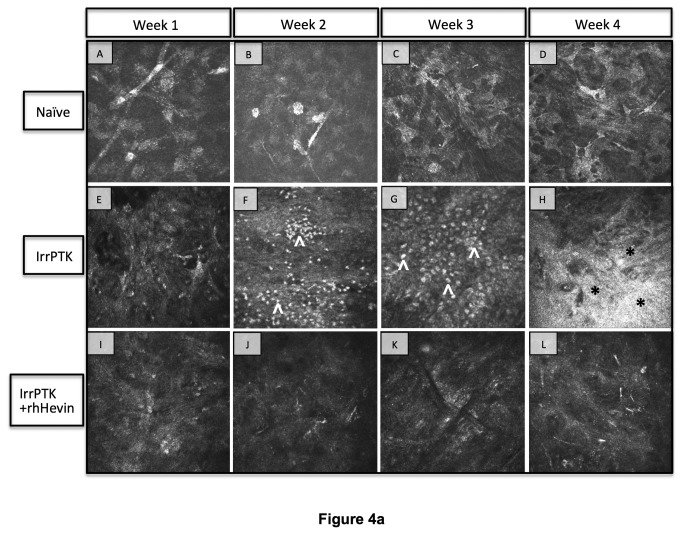
Figure 4. In vivo confocal microscopy in WT and Hevin-/- mouse.
Naïve WT (A-D) and Hevin-/- (A’-D’) mice corneas served as control samples and exhibit no cell infiltrates and stromal haze. IrrPTK-surgery triggered infiltration of inflammatory cells (^) at 2 weeks post-op in WT mice (F), which subsides by week 4 (H). These inflammatory events are followed by the development of corneal haze (*) as seen in week 4 samples (H). In Hevin-/- mouse, inflammatory cells are seen as early as week 1 post-op (E’), which continued to increase up to week 4 (F’-H’). Supplementation of exogenous rhHevin decreased cell infiltration and impeded stromal haze formation in WT (I-L) and Hevin-/- mice (I’-L’), completely eliminated inflammatory cells in WT corneas (I-L) though low numbers of inflammatory cells were still observed in Hevin-/- mouse (I’-L’).

