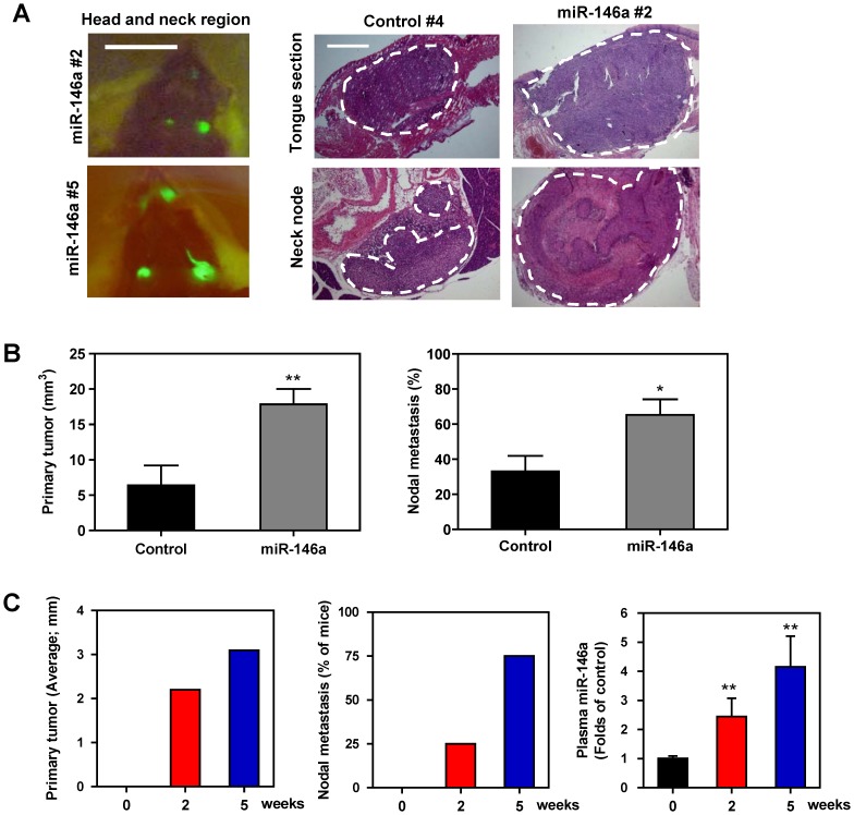
Figure 4. Exogenous miR-146a expression enhances the orthotopic tumorigenesis and neck nodal metastasis of SAS cells.
(A, B) Orthotopic tongue tumorigenesis and neck nodal metastasis of SAS cell subclones using the NOD-SCID mouse. (A) Lt, fluorescence images of head and neck region. Fluorescent primary tongue tumors and neck metastatic lesions are noted in vivo prior to autopsy. Middle panels, representative histopathological sections of the tongue and neck nodes. Bars, 1 cm or 1 mm (in Rt panels). The dotted lines delineate the lesions. Magnification of the histopathological sections, x25. (B) Quantification. Lt, the volume of primary tumors; Rt, the incidence of nodal metastasis. Exogenous miR-146a expression is significantly associated with increased burden of primary tumor burden and neck metastasis of mice. Data shown are the means ± SE from at least six mice. ns, not significant; *, p<0.05; **, p<0.01; Mann-Whitney test. (C) Quantification of the diameter of orthotopic tumors (Lt); the percentage of mice exhibiting visible or palpable neck masses (Middle); the plasma miR-146a (Rt) at different time points following the orthotopic injection of the SAS cell subclone with exogenous miR-146a expression. Progressive increase of plasma miR-146a that occurs in parallel with tumor growth and neck metastasis is noted. Data shown are the means or means ± SE from four mice. **, p<0.01; Wilcoxon’s test.

