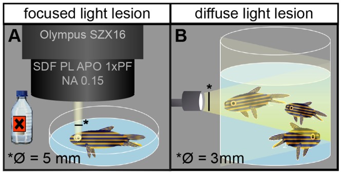
Figure 1. Schematic illustration of lesion paradigms.

A: In focused light lesion the fish is anaesthetized under the stereoscope and exposed to light onto one eye from one single angle, producing a well circumscribed lesioned area in the illuminated retina, next to non-illuminated control areas. The non-illuminated eye serves as an additional internal control. B: In diffuse light lesions, fish swim freely in a beaker and are exposed to light from all angles and to both eyes.
