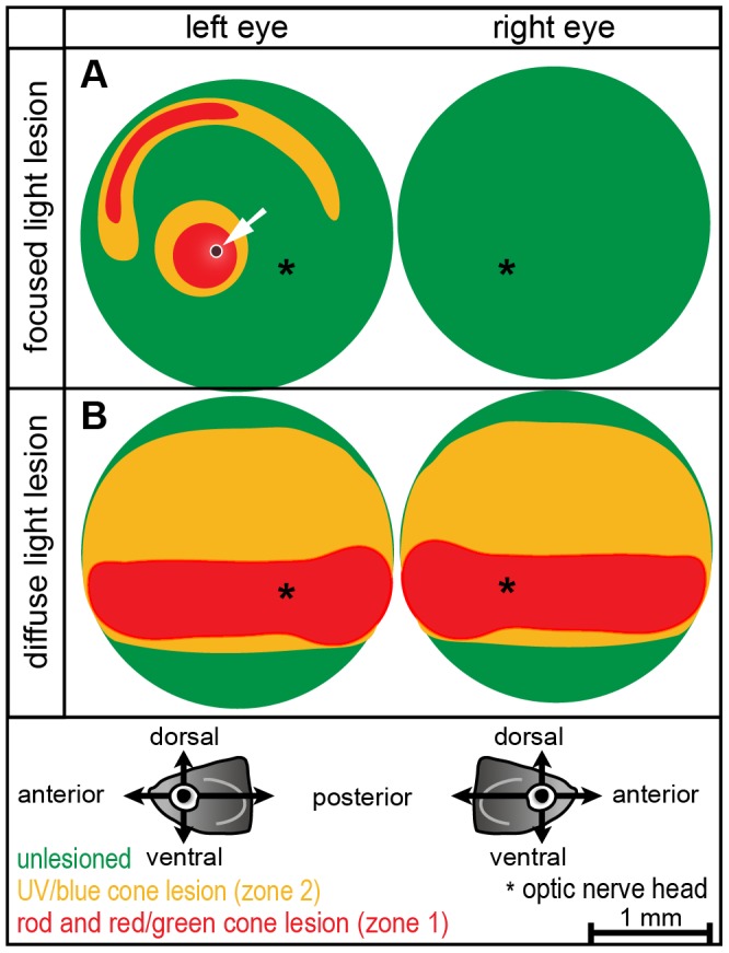
Figure 4. Schematic illustration of lesion patterns.

A: Focused light lesions form a complex lesion pattern in the central and peripheral retina. A small area of unspecific damage in all retinal layers is due to the high intensity of the center beam, and is indicated by the white arrow. Most of the light treated retina and the control eye on the other side is undamaged (green). B: Diffuse light lesions in the central retina are shaped as horizontal stripe. Around the area in the center (depleted of all photoreceptors) are UV and blue cone depleted areas in the peripheral retina (yellow). The ventral part is never damaged in any fish. (Orientation as shown in illustrations below; asterisk represents optic nerve head).
