
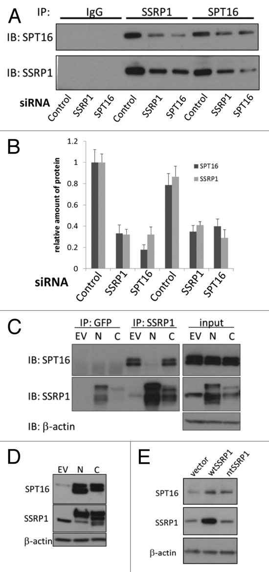
Figure 6. The stabilizing effect of SSRP1 mRNA on SPT16 protein levels does not require direct interaction of the two proteins. (A and B) Reduction of mRNAs of SSRP1 and SPT16 leads to stoichiometric reduction of both proteins in the complex.(A)Western blotting of immunoprecipitates of SSRP1 and SPT16 or control IgG in HeLa cells transfected with the indicated siRNAs.(B)Quantitation of data shown in (A). Band intensities of SSRP1 and SPT16 in control siRNA-transfected cells immunoprecipitated with anti-SSRP1 antibodies were set as 1.0.(C)HeLa cells were transfected with the empty EGFP fusion vector (no protein fused to EGFP, EV), or with constructs producing SSRP1 with EGFP fused to either its N or C terminus (N and C, respectively). At 48 h after transfection, extracts of the cells were used for immunoprecipitation with either GFP or SSRP1 antibodies. Immunoprecipitates were immunoblotted with antibodies, shown on the right. GFP-C-term-SSRP1 was not immunoprecipitated with anti-GFP antibodies (in multiple experiments), although it was precipitated with antiSSRP1. GFP is also visible in these cells.(D)Populations of HeLa cells transduced with the constructs indicated at the top of the figure were expanded and sorted for GFP expression. After establishing and expanding ~100% GFP-positive cell populations (~2 wk), cell extracts were analyzed by western blotting to assess expression of exogenous SSRP1 variants and associated changes in SPT16 protein levels. Note that it was difficult to obtain a stable cell population overexpressing SSRP1 constructs, including wild type SSRP1 (see “Discussion”). However, in all cases in which SSRP1 protein expression was increased after cell expansion, SPT16 protein levels were found to be increased as well. (E) Overexpression of a mutant SSRP1 mRNA that cannot be translated leads to increased SPT16 protein levels. WI38 cells were transduced with either an empty expression vector (vector), or vectors directing expression of the wild type SSRP1 mRNA (wt) or a mutant SSRP1 mRNA with a stop codon inserted after the first methionine (nt). After puromycin selection and expansion of transduced cells, total cell extracts were analyzed by western blotting with the indicated antibodies.