
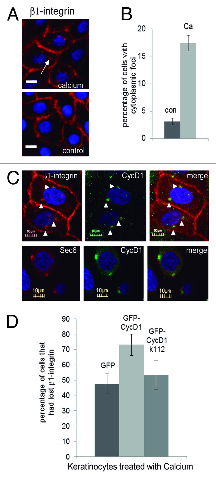
Figure 5. Co-localization of CycD1 with β1 integrin during keratinocyte differentiation. Before fixation, keratinocytes were incubated for only 16 h in the presence of 2 mM calcium to look at cells at the initial steps of differentiation. (A) β1 integrin was analyzed by immunofluorescence. Images were acquired by using a 40× objective in an Olympus IX71 inverted microscope (20 µm bar) (B) Quantification of cells with β1 integrin in cytoplasmic foci. Data are expressed as mean ± SEM from two independent experiments (P = 0.045; n ≥ 122). (C) Co-localization of CycD1 with β1 integrin and Sec6 was analyzed by confocal microscopy using a 60× objective (10 μm bar). Nuclei were stained with Hoescht. (D) Keratinocytes were transfected with either GFP-CycD1, GFP-CycD1k112, or GFP and after a short time of incubation with calcium, cells were fixed and processed for IF to detect β1 integrin, and the percentage of cells that had completely lost β1 integrin signal in the membrane was counted. Percentage of cells and confidence limits (α = 0.05; n ≥ 110) are plotted.