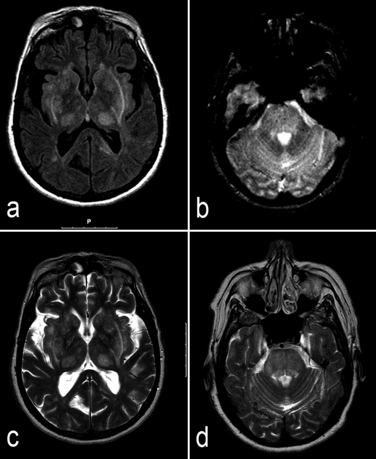
Figure 1.
MRI scans: admission MRI scan axial fluid-attenuated inversion recovery (A) and T2* (B) images showing symmetrical lesions evolving the basal ganglia, thalamus, internal and external capsules and brain stem, with expansion and microhaemorrages in the pons. Follow-up (7 months later) MRI scans: axial T2 images (C and D) showing partial resolution of the admission lesions.

