Figure 1.
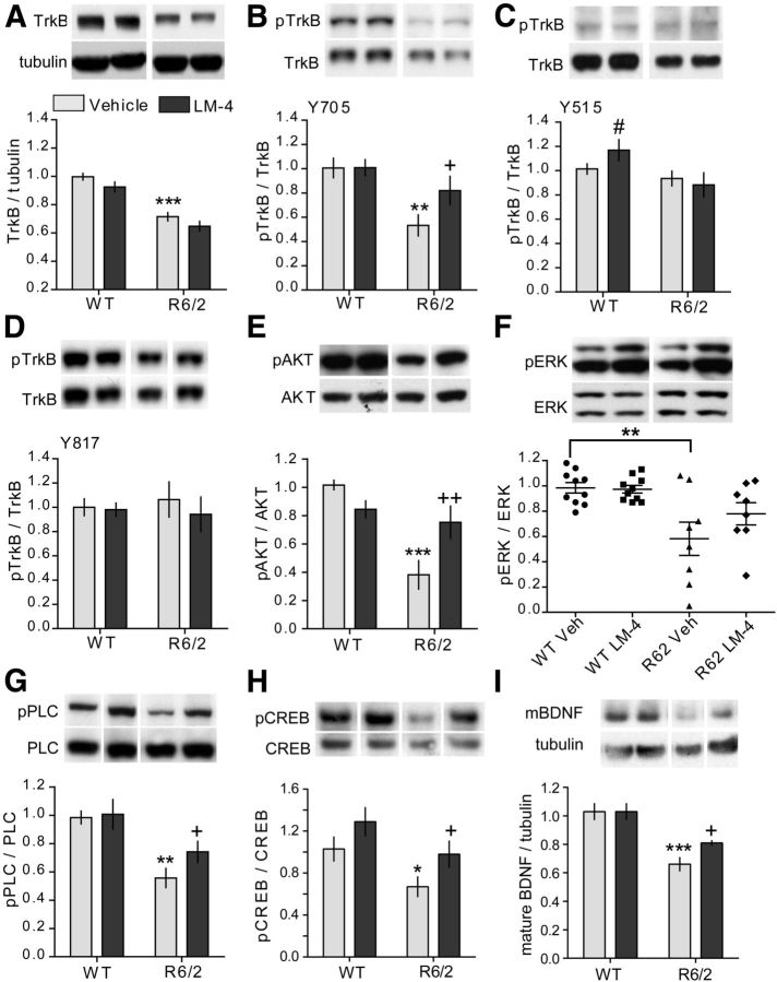
LM22A-4 activates TrkB and its associated downstream signaling in striatum of R6/2 mice. Representative Western blots of striatal homogenates from 11- to 12-week-old WT or R6/2 mice treated with vehicle (Veh) or LM22A-4 (LM-4) for 7 weeks. For all quantitation, n = 8–10 mice/group were used and values were normalized to the WT Veh group run on the same gel. Immunobands from one mouse per group and corresponding densitometric group analyses are shown. A–D, Full-length TrkB levels (A) and its phosphorylation (p) at tyrosine residues Y705 (B), Y515 (C), and Y817 (D) are depicted. Samples from each mouse were run in three to four replicates. TrkB levels were lower in striatum of R6/2 mice relative to WTs (***p ≤ 0.001 vs WT Veh) regardless of LM-4 treatment. TrkB phosphorylation (pTrkB) at Y705 (**p ≤ 0.01 vs WT Veh), but not Y515 or Y817, was decreased in Veh-treated R6/2 mice compared with WTs. LM-4 inhibited the reduction in Y705 phosphorylation (+p ≤ 0.05 vs R6/2 Veh). LM-4 also increased phosphorylation at Y515 in WT mice (#p ≤ 0.05 vs WT Veh; two-tailed Student's t test). E–G, Western blots showing phosphorylation of AKT (E), ERK (F), and PLCγ (G), the three main downstream signaling pathways of TrkB. Samples from each mouse were run in four to six replicates. Phosphorylation of AKT, PLCγ, and ERK was decreased in Veh-treated R6/2 mice compared with WTs (**p ≤ 0.01 and ***p ≤ 0.001 vs WT Veh). LM-4 prevented the deficits in AKT and PLCγ (+p ≤ 0.05 and ++p ≤ 0.01 vs R6/2 Veh) but not ERK phosphorylation. H, Western blot showing phosphorylation of CREB at Ser133. Samples from each mouse were run in duplicate. pCREB was reduced by 36 ± 10% in R6/2 mice given Veh (*p < 0.05 vs WT Veh; two-tailed Student's t test) and LM-4 prevented this decrease (+p < 0.05 vs R6/2 Veh; two-tailed Student's t test); there was also an increase in pCREB in the WT LM-4 group that did not reach statistical significance (p = 0.07 vs WT Veh). I, Western blot showing mBDNF protein levels in striatum. Samples from each mouse were run in triplicate. mBDNF levels are reduced in striatum of R6/2 Veh mice (***p ≤ 0.001 vs WT Veh) and this difference was attenuated by LM-4 treatment (+p ≤ 0.05 vs R6/2 Veh; two-tailed Student's t test). For all analyses, results are expressed as the mean ± SEM and, for comparison purposes, some nonadjacent lanes of Western blots were moved together and separated by spaces. Statistical significance was determined by ANOVA with Newman–Keuls post hoc testing, unless otherwise noted.

