Figure 1.
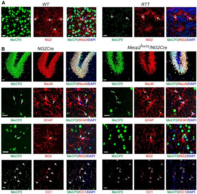
MeCP2 protein is specifically depleted from the oligodendrocyte lineage cells in brains of Mecp2loxJ/y/NG2Cre mice. A, Representative images of brain sections of WT and RTT (Mecp2J.-/y) (Chen et al., 2001) male mice coimmunolabeled for MeCP2 (green) and NG2 (red). DAPI (blue) represents nuclear staining. MeCP2 (green) is expressed in OPCs (NG2+, red) in the hippocampus of WT mice and absent in RTT mice. Scale bars, 20 μm. B, Representative images of brain sections showing the presence of MeCP2 in all cell types in the brain of NG2Cre male mice and its specific absence in the oligodendrocyte lineage cells (NG2+ and CC1+ cells) in the brain of MeCP2loxJ/y/NG2Cre male mice. Sections were coimmunolabeled for MeCP2 (green) and neuronal marker (NeuN, red) in the dentate gyrus area, MeCP2, and astrocytic (GFAP, red) or OPCs (NG2, red) markers in the hippocampus, and MeCP2 and mature oligodendrocytic marker (CC1, red) in the corpus callosum. DAPI (blue) represents nuclear staining. Scale bars, 20 μm. Arrows indicate colocalization or absence of MeCP2 in GFAP, NG2, or CC1-positive cells.

