Figure 7.
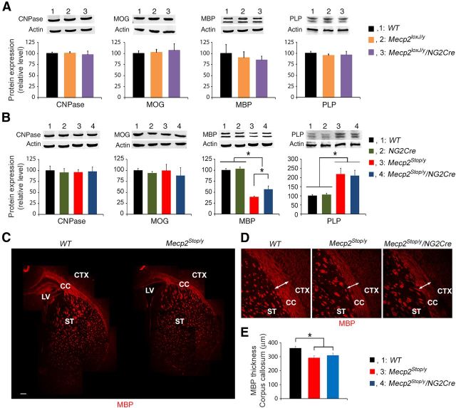
MeCP2 expression in the oligodendrocyte lineage cells only partially rescues the aberrant expression pattern of MBP protein in RTT mice. A, The loss of MeCP2 only in the oligodendrocyte lineage does not affect the expression pattern of proteins involved in myelination. Quantitative Western blot analysis showing the expression levels of proteins involved in myelin formation in whole-brain extracts of P16 Mecp2loxJ/y/NG2Cre and the indicated control mice. B, Expression of MeCP2 in Mecp2Stop/y/NG2Cre mice only partially restored the reduced expression of MBP observed in Mecp2Stop/y mice. Quantitative Western blot analysis showing aberrant expression of MBP and PLP in whole-brain extracts of P16 Mecp2Stop/y mice and partial restoration of MBP expression in Mecp2Stop/y/NG2Cre age-matched littermate mice. C, Reconstruction of MBP confocal images of the corpus callosum (CC) and the striatum (ST) areas of the WT and Mecp2Stop/y brains. CTX, Cortex; LV, lateral ventricle. Scale bars, 200 μm. D, High magnification of the MBP immunostaining in the corpus callosum areas. Arrows indicate the thickness of MBP staining in the corpus callosum area calculated in E. E, Bar graphs representing the thickness of MBP staining in the corpus callosum area of the WT, Mecp2Stop/y, and Mecp2Stop/y/NG2Cre brains. An average of three measurements along the corpus callosum for each image was used for the thickness calculation. One-way ANOVA followed by Holm-Sidak post hoc for multiple-comparisons test was used to determine differences between groups in A, B and E. Error bars indicate mean ± SEM. *p < 0.05. n = 3 mice per genotype.

