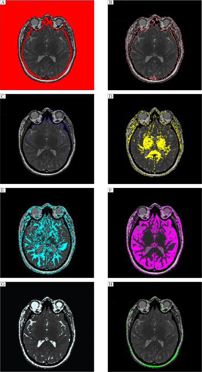
Fig. 6. Axial slices with only one range of resistivity colored against a combined gray image to illustrate the types of tissues that are included for each range of resistivity.
Note that there is some overlap between tissue types. A = cortical bone and air, B = soft tissue, C = dura and vessel walls, D = white matter, F = grey matter, G = spinal fluid, H = subdermal skin.

