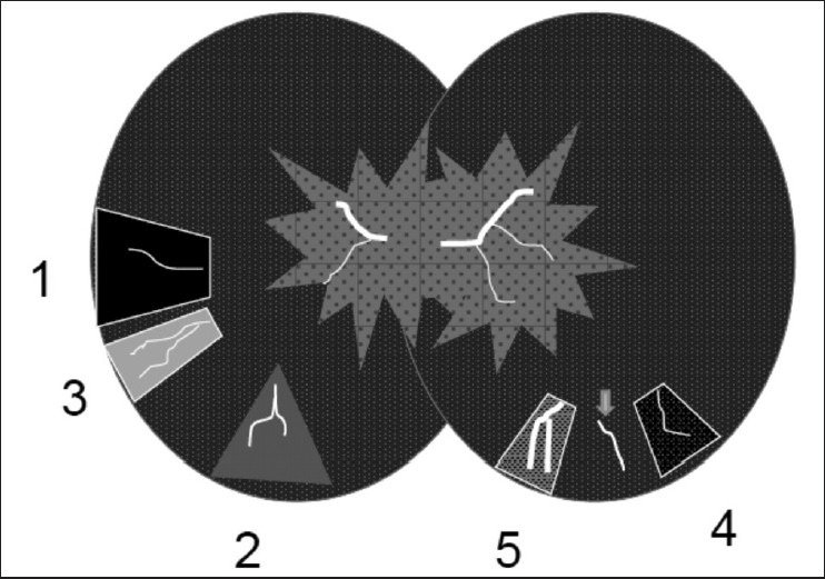
Figure 13.

Schematic model of heterogeneous lung attenuation. Airway disease (left) can lead to (1) gas trapping and reflex vasoconstriction seen as hypoattenuating regions with fewer or thinner blood vessels. (2) Diversion of blood flow to normal lung can make it hyperattenuating. (3) Complete loss of ventilation can lead to atelectasis, crowding of blood vessels, and hyperattenuation. Vascular disease can also lead to heterogeneous lungs (right) due to (4) increased vascular resistance and hypoperfusion (e.g., pulmonary hypertension) that contrasts with normal lungs and vascularity (arrow) and (5) regions of increased perfusion
