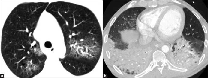
Figure 6.

(a) GGO seen as increased attenuation of the lung parenchyma on HRCT without obscuring the underlying vascular or bronchial margins. (b) Consolidation in a case of aspiration pneumonia presents as homogeneous increased parenchymal attenuation in the lower lobes obscuring the vascular and bronchial margins with air bronchogram
