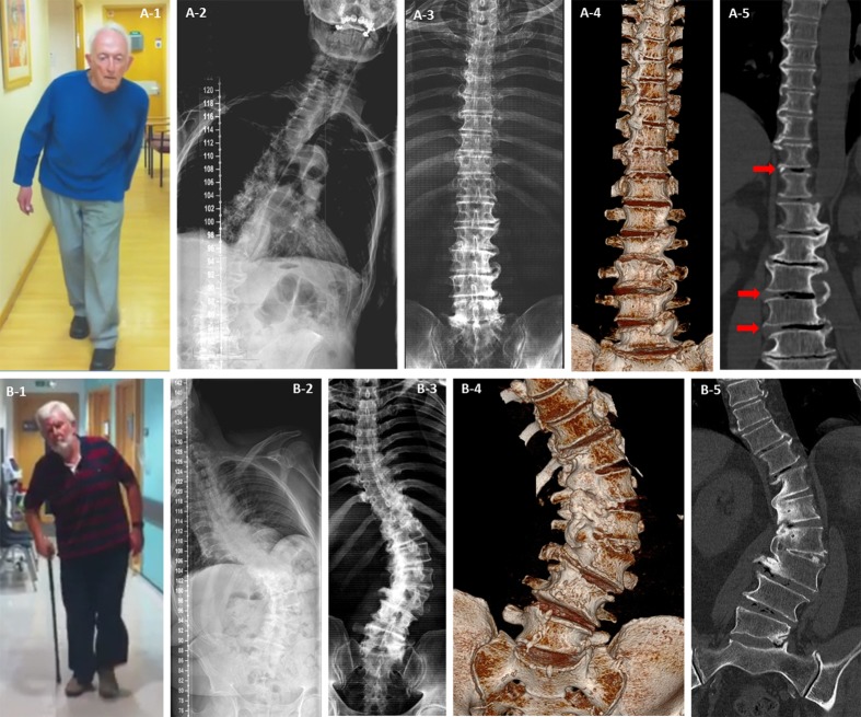
Figure 1.
Mobile and fixed scoliosis in Pisa syndrome. Patient A had scoliosis on standing radiograph (A-2) but not when he was scanned supine (A-3, A-4 and A-5). There was evidence of osteophytic overgrowth below the apex of the scoliosis in the lumbar spine and above on the opposite side in the thoracic spine (A-4 and A-5), this pattern suggests the degenerative changes were working to stabilise his spine but stopped short at the apex of his curve leaving him mobile but tilted at that level when standing (A-1 and A-2). The reduction in curve with position, presence of interdiscal gas (red arrows A-5) and gaps between the osteophytes are evidence that despite attempts the deformity is not fixed. Patient B had only minor improvement of his scoliosis on supine positioning (9% reducibility) (B-2 and B-3). Fusion of vertebral segments due to complete osteophytic bridging at the apex of the curve was clearly seen (B-4 and B-5) resulting in a fixed and possibly stable spinal deformity. Key: 1=patient photographs of Pisa syndrome while walking; 2=standing full spine anterior–posterior radiograph; 3=supine CT scan two-dimensional composite image; 4=supine CT scan three-dimensional surface rendered image; 5=supine CT scan two-dimensional fine cut in coronal plane.

