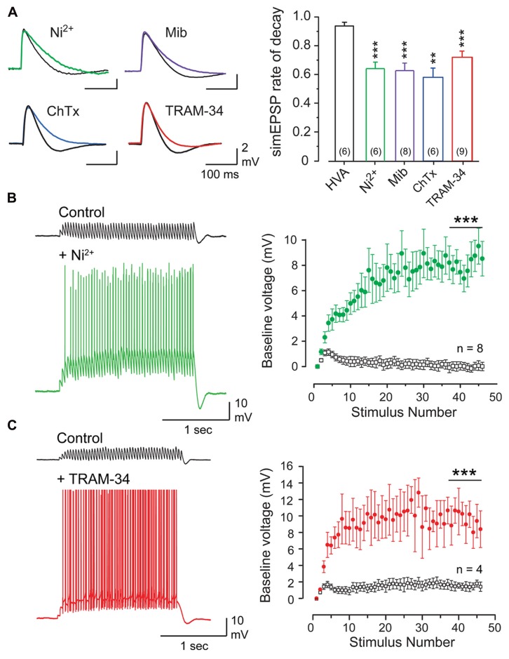
FIGURE 3.
The parallel fiber EPSP activates a Cav3- and KCa3. 1-mediated AHP that suppresses temporal summation. (A) Superimposed records of simEPSPs in Purkinje cells before and after 100 μM Ni2+, 1 μM mibefradil (Mib), 100 nM ChTx, or 100 nM TRAM-34. Bar graphs show mean values of the reduction of simEPSP rate of decay. HVA refers to a cocktail of ω-conotoxin GVIA (1 μM), nifedipine (1 μM), and SNX-482 (200 nM). (B,C) Recordings and plots of the mean baseline voltage during 25 Hz trains of parallel fiber-evoked EPSPs before and after applying Ni2+ [(B), 100 μM)] or TRAM-34 [(C), 100 nM]. Recordings in (B,C) were conducted in 50 μM picrotoxin. Statistical significance tested for last 10 pulses of stimulus trains in (B,C) is denoted by bars. Sample values in (A) are shown in brackets within bar plots. **p < 0.01, ***p < 0.001. Modified from Engbers et al. (2012).

