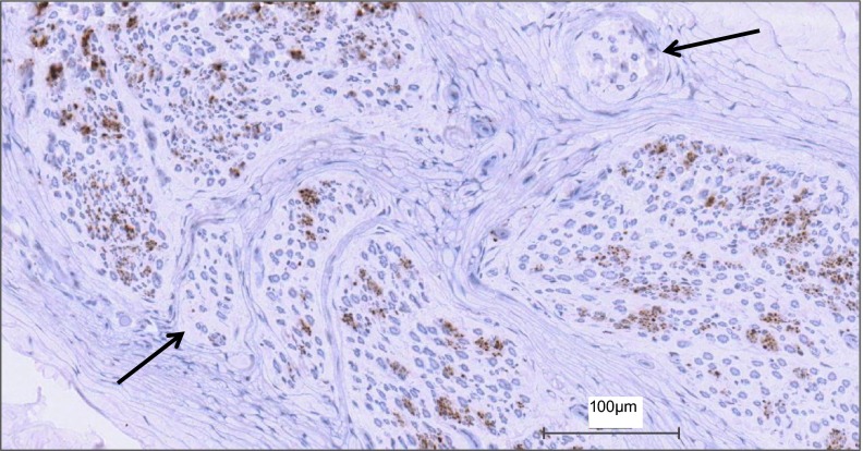
Fig. 2.
View of how sympathetic fibers (positively stained for tyrosine hydroxylase) vary with each fascicle in a representative section of the common peroneal nerve. The amount of staining within a fascicle varied between fascicles of the same nerve. Some fascicles displayed no staining, while others showed a large abundance. Black arrows indicate fascicles with no sympathetic axons.

