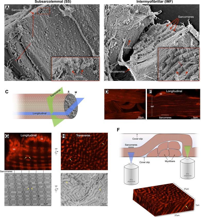
Fig. 2.
Topological and morphological differences between subsarcolemmal (SS) and intermyofibrillar (IMF) mitochondria. A: scanning electron microscopy (SEM) imaging of a freeze-fractured soleus muscle fiber sectioned in the cross-sectional plane with its cytoplasm exposed. The SS region is outlined where mostly globular SS mitochondria are clustered. B: SEM of freeze-fractured muscle fiber cracked to expose the staircase-like sarcomere structure revealing the IMF mitochondrial reticulum between each sarcomeric plane. C: diagram representing the two main planes—longitudinal and transverse (i.e., cross-section)—used to quantify various aspects of mitochondrial morphology. D and E: confocal imaging of permeabilized muscle fibers labeled with Mitotracker Red showing SS and IMF mitochondria and sarcomeric organization. F: schematic of experimental setting used for optical sectioning of Mitotracker-labeled muscle fibers in the longitudinal (blue) and cross-sectional (green) planes. Below is a three-dimensional reconstruction of an oblique section across a myofiber, showing elongated organelles (arrow). G: the longitudinal view in both confocal and electron microscopy reveals the pairwise arrangement of IMF mitochondria across Z-lines at each sarcomeric plane (appearing as spherical organelles, arrows), whereas the transverse view reveals the actual tubular morphology of IMF mitochondria (H, arrows).

