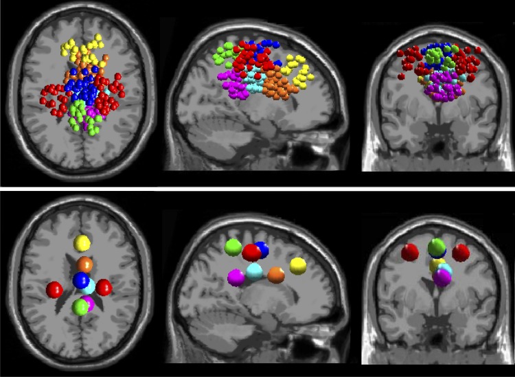
Fig. 2.
Clusters of independent component (IC) EEG sources localized in and near anterior cingulate (orange), posterior cingulate (2 clusters, magenta and cyan), superior dorsolateral-prefrontal (yellow), anterior parietal (green), left and right lateral sensorimotor (red), and medial sensorimotor (blue) cortex. Top: small spheres indicate the equivalent current dipole locations of each clustered IC source. Bottom: larger spheres show the locations of the cluster centroids.

