
Figure 3.
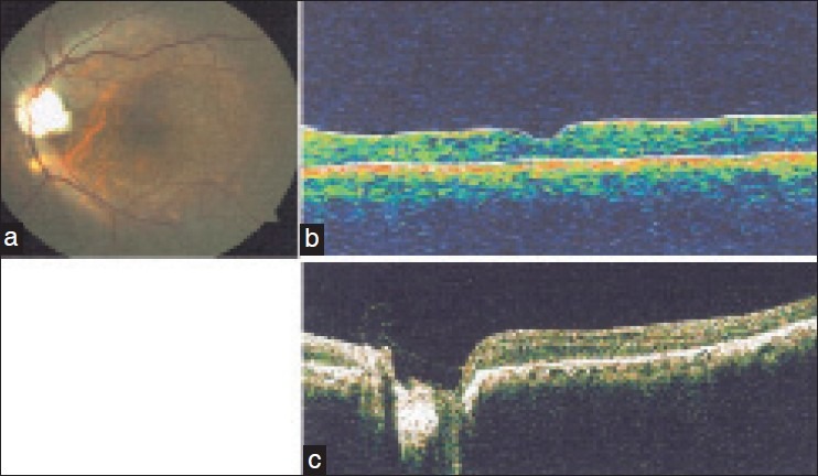
(a) Postoperative fundus photo with regressed macular detachment. (b) Postoperative optical coherence tomography (OCT) confirming flat macula. (c) Postoperative OCT confirming presence of inverted internal limiting membrane covering the optic disc pit