
Figure 2.
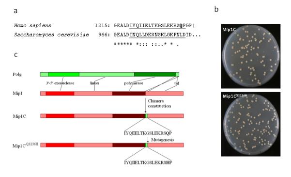
(a) Alignment of C-terminal stretch of human polγ and the corresponding stretch of yeast Mip1. Q1236 amino acid is in bold; the region which is changed in Mip1C is underscored. (b) Linear representation of polγ, Mip1, Mip1C and Mip1CQ1236H organization. Q1236 amino acid is in bold. (c) Petri dish images showing the normal and petite colonies from the parental (Mip1C) and the Mip1CQ1236H strain (bottom).