
Figure 3.
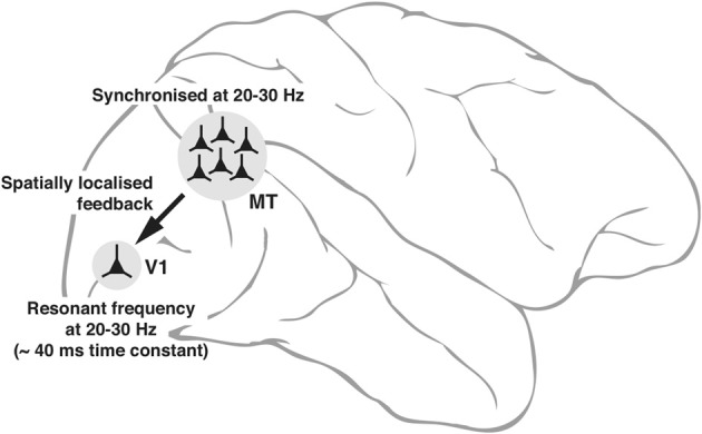
Schematic diagram, explaining the neuronal basis of feedback oscillations. Local network properties and biophysical parameters such as time constants of neurons determine the synchronizing frequencies of neurons in MT that provide feedback to V1 and the resonant properties of neurons in V1. It is suggested that a group of neurons representing a single location (of say, an object) in the parietal saliency map sending its signals via the corresponding group of neurons in MT can set the membrane potential of the spatially equivalent group of neurons in V1 (only 1 shown) to oscillate at its resonant frequency. Such an oscillation would facilitate the V1 neurons to respond to a sensory input more readily as the input signals ride on the top of the depolarizing crests in membrane potential.