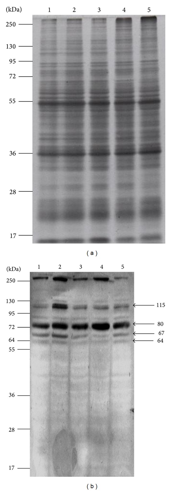
Figure 1.

Midgut total protein extraction with Triton X-100 and VOPBA. (a) Proteins were extracted from mosquito MG tissue at different Triton X-100 concentrations, separated by SDS-PAGE, and stained with Coomasie Blue. Triton X-100 concentrations were 0.01, 0.05, 0.1, 0.5, and 1% corresponding to lane 1 to 5, respectively. (b) Proteins, separated by SDS-PAGE, were blotted onto PVDF and incubated with biotinylated DENV-2 and then with AP-Streptavidin. Proteins recognized by DENV-2 were developed with BCIP/NBT according to the procedure previously described [8]. The apparent molecular weights of these proteins are shown on the right side of (b). Molecular weight markers are shown on the left side in (a) and (b).
