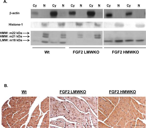
Figure 3.
(A) Representative Western blot of FGF2 isoform localization in non-ischemic wildtype (Wt), FGF2 LMWKO and FGF2 HMWKO mouse hearts. In Wt hearts, the LMW, 18 kDa isoform was localized to the cytosolic (Cy) and nuclear (N) fractions of the heart; whereas, the HMW, 21 and 22 kDa, isoforms were nuclear localized. In the absence of the LMW isoform (FGF2 LMWKO), the HMW isoforms were only expressed in the nucleus. In the absence of the HMW isoforms (FGF2 HMWKO), the LMW isoform was localized in the cytoplasm and nucleus of the heart. β-actin, a cytosolic protein, used as a measure of cytosolic fraction enrichment. Histone-1, a nuclear protein, used as a measure of nuclear fraction enrichment. (B) Localization of FGF2 isoforms via immunohistochemistry in adult mouse ventricular myocardium. Wildtype (Wt) heart (first panel), FGF2 LMW KO heart (middle panel), and FGF2 HMWKO heart (last panel). Brown color indicates FGF2, and blue color marks nuclei that are counterstained by hematoxylin. In wildtype and FGF2 HMWKO (expression of only LMW isoform) hearts, FGF2 is found in both cytoplasm and nucleus of the cardiomyocytes. FGF2 is detected in FGF2 LMW KO (expression of only HMW isoforms) hearts. Scale bar in A–C, 50 μm. n=3/group.

