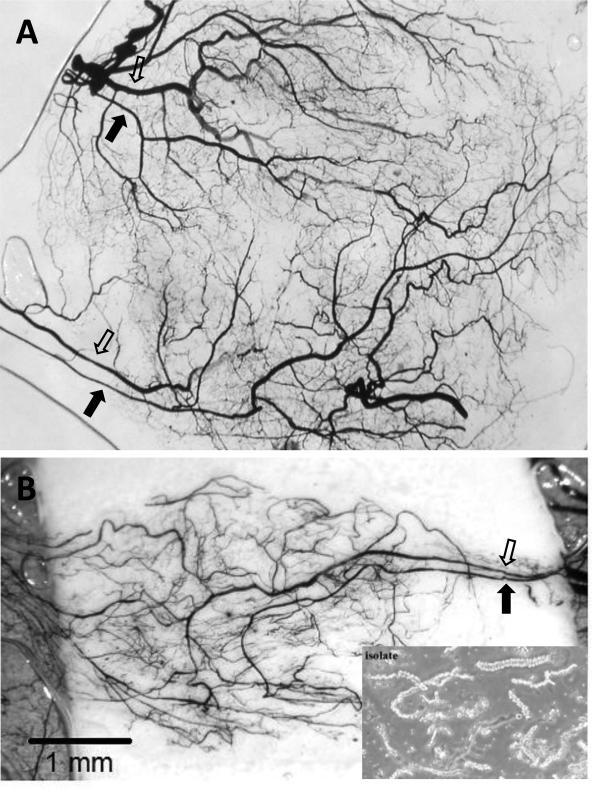
Figure 2.
Two separate examples (A and B) of regenerated microvascular beds derived from implanted isolated microvessels visualized by india ink casting and tissue clearing. In both examples, paired inflow (closed arrow) and outflow (open arrow) pathways developed a branched microvascular tree. Notice that in (A), two “microvascular units” evolved from the initial implanted parent microvessel source (lower right inset). Because the microvasculatures derived from these implants progress in a regimented fashion in the absence of additional stromal cells, they are useful for investigating angiogenesis and post-angiogenesis processes and mechanisms. The implants consisted of isolated, intact microvessel segments suspended in polymerized, 3-dimensional, type I collagen gels and implanted subcutaneously for 4 weeks in mice.

