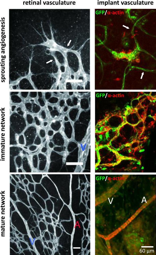
Figure 4.
Vascular and network morphologies present during progression through neovascularization in two different settings: post-natal development of the retinal vasculature and regenerative implant neovascularization. Initial angiogenic sprouts (white arrows) inosculate to form an immature network which matures into a hierarchical tree of mature arterioles (A), venules (V) and capillaries. Scale bars in the left panels equal 50 μm. Parts of this figure were derived from [70] and [172] with permission.

