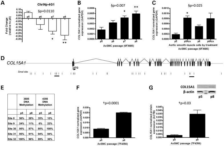
Figure 2.
Identification of COL15A1 as a gene that is epigenetically regulated in smooth muscle replicative aging. (A) The clone containing COL15A1 exhibits decreased methylation with passage. This was assessed by microarray hybridization of p5, p6, p7 and p8 methylated DNA and displayed as fold change (log2[p8/p5]). (B) COL15A1 gene expression was measured by RT-PCR with age of SMC (n = 3) and normalized to glyceraldehyde-3-phosphate dehydrogenase (GAPDH). (C) Normalized COL15A1 gene expression was measured by RT-PCR in both young and aged cells in the absence and presence of a demethylating chemical, 5-Aza-2′-deoxycytidine (Aza). Data were normalized to GAPDH. (D) A schematic of the clone containing COL15A1 identified in a screen for genes that change DNA methylation with SMC age. The start codon (ATG) is contained within the first exon; exons are denoted as black discs and introns as lines. SmaI sites contained within this region are denoted by a black vertical line. Groups of SmaI sites that are hypomethylated with age are underlined. (E) Bisulfite cloned sequencing results from two passaged AoSMCs lines. Indicated sites are part of the two underlined clusters in the gene schematic. (F) Validation of COL15A1 expression changes with passage in second AoSMC line (7F4356). (G) Total cellular COL15A1 protein levels were assayed in young (p5) and aged (p8) SMCs by western blot. Quantitation (n = 3) was performed on a LI-COR Odyssey Infrared Imaging System. Data were normalized to β-actin (ACTB). A representative western blot is shown for COL15A1 and loading control β-actin. §P, repeated measures ANOVA, *Bonferroni's Multiple Comparison test P < 0.05; **P < 0.01. †P, paired t-test (n = 3), two-tailed.

