
Figure 2.
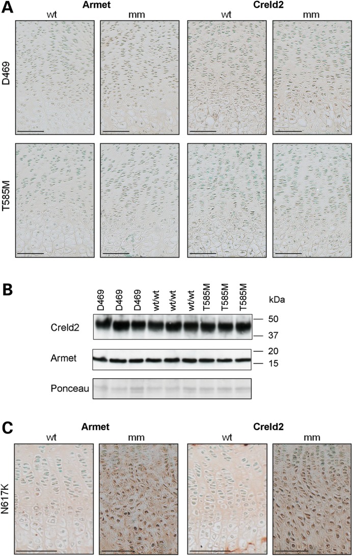
Creld2 and Armet are not up-regulated in mouse models of COMP-related PSACH-MED, but are increased in a model of MCDS. (A) IHC using Armet and Creld2 antibodies on tibia growth plates from 3-week-old Comp T585M (T585M), Comp D469del (D469) and matched wild-type (WT) mice. No increase in staining was observed for Armet or Creld2 in either mutant (mm) mouse model compared with wild-type (WT). (B) Chondrocytes were isolated from the cartilage of 5-day-old Comp T585M (T585M), Comp D469del (D469) and wild-type (WT) mice. Total protein from 1 × 10⁵ cells was loaded per lane and analysed by SDS–PAGE and western blotting. No detectable differences in the levels of Creld2 and Armet were observed in cell extracts from mutant mice (T585M and D469) compared with wild-type (WT) controls. Equal protein loading was verified by Ponceau staining. (C) IHC on tibia growth plates from 3-week-old Col10a1 N617K (N617K) mice demonstrated both intracellular and ECM staining in the hypertrophic zone of growth plate cartilage. Scale bar is 100 µm; kDa = kilodaltons.