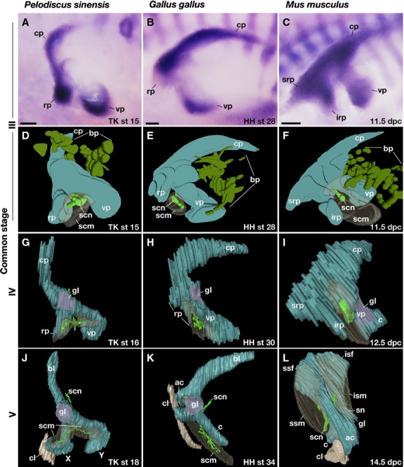
Fig. 2.

Comparison of the shoulder girdle development. (A,D,G,J) Pelodiscus sinensis, (B,E,H,K) chickens, and (C,F,I,L) mice. (A–C) Expression of Sox9 in the left girdle. (D–L) Three-dimensional reconstructions of the cartilage, nerves, and muscles. All panels are the lateral view, and rostral is to the left. bp, brachial plexus; c, coracoid; cl; clavicle; cp, caudal process; irp, infrarostral process; isf, infraspinatus fossa; ism, infraspinatus muscle; rp, rostral process; scm, supracoracoid muscle; scn, supracoracoid nerve; sn, scapular neck; srp, suprarostral process; ssf, supraspinatus fossa; ssm, supraspinatus muscle; vp, ventral process. Scale bar: 200 μm.
