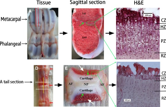
Fig. 1.

Specimen preparation. (A-C) Metacarpal joint. (A) Calf metacarpal-phalangeal joint. Red dashed lines indicate where specimen was sagittally dissected. (B) Sagittally dissected specimen, green arrow highlights the growth plate separating the primary (1st) and secondary (2nd) ossification centres. Green square highlights growth plate specimen dissected out for histology. (C) H&E stained micrograph of the metacarpal growth plate. (D-F) Vertebral joint. (D) Calf tail joints. The vertebral joint was dissected through the white dashed lines, then sagittally dissected along the yellow dashed line. Red arrows indicate the intervertebral discs. (E) A mid-sagittal dissected specimen, illustrating the general structure of a vertebral joint composed of the disc nucleus pulposus (NP) in the centre surrounded laterally by the disc annulus fibrosus (AF), longitudinally by cartilages which directly attached to the vertebral bone (white arrows) though the growth plate (green arrows). Green square highlights growth plate specimen dissected out for histology. (F) H&E stained micrograph of the vertebral growth plate. RZ, resting zone; PZ, proliferative zone; HZ, hypertrophic zone; CZ, calcified region of the hypertrophic zone.
