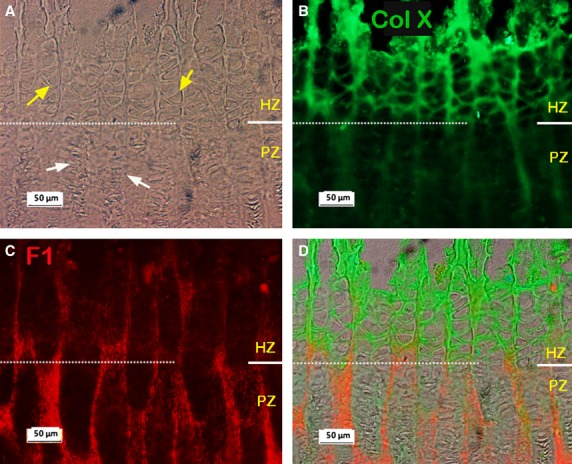
Fig. 3.

Typical dual-immunostained collagen X (Col X; green) and fibrillin-1 (F1; red) in the metacarpal growth plate. (A-C) Micrographs from same site, white dashed line separates the HZ and PZ regions. (A) Phase contrast image. (B) Immunostained collagen X (Col X) showing strong staining for Col X in the HZ region. (C) Immunostained F1 showing relatively more F1 in the PZ than in HZ regions. (D) Merged image of A, B and C. RZ, resting zone; PZ, proliferative zone; HZ, hypertrophic zone.
