Fig. 4.

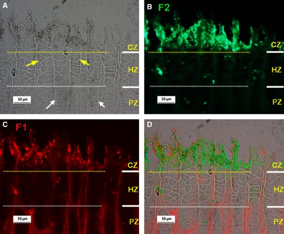
Typical dual-immunostained fibrillin-1 (F1; red) and fibrillin-2 (F2; green) in the metacarpal growth plate (pre-treatment condition: hyaluronidase 2hrs at 37°C; longer incubation in hyaluronidase can lead to loosening and loss of the calcified tissue). (A-C) Micrographs from the same site, yellow and white dashed lines separating the CZ from the HZ and the HZ from the PZ regions respectively. (A) Phase contrast image, white and yellow arrows indicate typical proliferative and hypertrophic cell columns respectively. (B-C) Immunostained F2 and F1 respectively, showing little F2 staining is visible in the HZ and PZ regions but strong staining for F2 is apparent in the CZ where F1 is also apparent. (D) Merged images of A-C. RZ, resting zone; PZ, proliferative zone; HZ, hypertrophic zone; CZ, calcified region of the hypertrophic zone.
