
Figure 6.
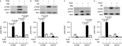
PI3K-dependent and PDK1-independent Akt1 phosphorylation and PI3K-/PDK1-dependent Akt2 phosphorylation under cell-free conditions. Akt1 or Akt2 was reacted with (+) and without (−) PI3K (1 μg/ml) (A and C) or PDK1 (1 μg/ml) (B and D) in the presence and absence of wortmannin (WM) (20 nM) or BX912 (BX) (100 nM), and western blotting was carried out using antibodies against pT308(9), pS473(4), and Akt1/2. Signal intensities for phosphorylated Akt1 (pAkt1) or Akt2 (pAkt2) were normalized to those for Akt1 or Akt2. In the graphs, each value represents the mean (±s.e.m.) intensity for pAkt1 or pAkt2 at each site (n=4). P values, Dunnett's test. NS, not significant.