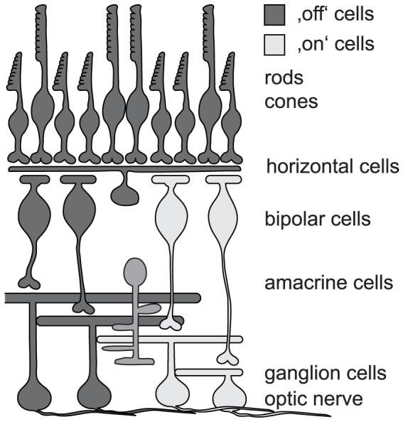
Figure 1. Scheme of the mammalian retina.

Photoreceptors (rods and cones) hyperpolarize to light. Consequently, a successful vision restoration approach that targeted cones has utilized halorhodopsin as optogenetic tool [13]. As this strategy is using the earliest possible neurons within the retinal circuit it is most likely best suited to recreate the most meaningful (i.e. natural) light responses. There are two broad categories of bipolar cells: ‘on’ bipolar cells depolarize in response to light. They have successfully been targeted with ChR-2 to achieve vision restoration [16]. ‘Off’ bipolar cells hyperpolarize to light. Currently, there is no known promoter to drive optogene expression specifically in this cell group. Amacrine cells are a very diverse group of inhibitory interneurons. In particular A-II amacrine cells are discussed as a promising target for optogenetic intervention, as they hook into both ON and OFF circuitry in the retina. The discussion in the manuscript about targeting bipolar cells equally applies to A-II amacrine cells. Ganglion cells are the output neurons of the retina; their axons form the optic nerve. In terms of restoration of retinal processing (not just restoring light sensitivity) they are the least favored candidates for optogenetic intervention. In addition, targeting presynaptic neurons increases overall light sensitivity of the system by pooling of presynaptic input. Nevertheless, optogenetic vision restoration has successfully been performed with ganglion cells as targets [17].
