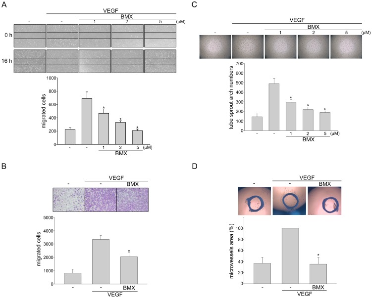
Figure 3. BMX inhibited VEGF-induced migration, tube formation and microvessel sprouting.
(A) HUVECs were starved in 2% FBS containing medium without ECGS for 16 h. Cell monolayer were then scratched and treated with vehicle or indicated concentrations of BMX in the presence of VEGF for another 16 h. The number of migrated cells was then determined. Each column represents the mean ± S.E.M. of four independent experiments. *p<0.05, compared with the group treated with VEGF alone. (B) After starvation as described in (A), Cells were then seeded in the top chamber in the absence or presence of BMX (5 µM) using VEGF as chemo-attractant. After 16 h, invaded cells through the gelatin-coated membrane were stained and quantified. Each column represents the mean ± S.E.M. of four independent experiments. *p<0.05, compared with the group treated with VEGF alone. (C) HUVECs were seeded on Matrigel in the presence of VEGF (20 ng/ml) with or without BMX at indicated concentrations. Cells were then photographed under phase-contrast microscopy after 16 h. Bar graphs show compiled data of average sprout arch numbers (n = 4). *p<0.05, compared with the group treated with VEGF alone. (D) Rat aortic rings were placed in Matrigel and treated with VEGF (20 ng/ml) in the presence or absence of BMX (5 µM). The effect of BMX on formation of vessel sprout from various aorta samples was examined on day 8. Bar graphs show compiled data of average microvessels area (n = 4). *p<0.05, compared with the group treated with VEGF alone.

