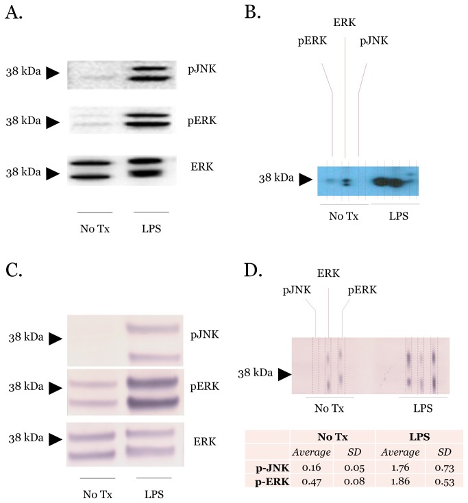
Figure 4. Protein immunoblots of the MAPK pathway using different chemiluminescent detection modalities.
(A) Traditional immunoblots of RAW264.7 cell lysates with or without LPS for 45 minutes. Membranes were probed for phospho-JNK and phospho-ERK using total ERK as the loading control. Primary antibodies were detected using HRP chemiluminescent techniques. (B) Microfluidic immunoblots mirroring conditions in (A). (C) and (D) are identical to (A) and (B) except they were performed using AP chemiluminescent secondary antibodies to detect primary antibodies. The signal intensities from three microfluidic immunoblots were quantified using ImageJ. Immunoblots are representative of at least three independent PVDF membranes.

