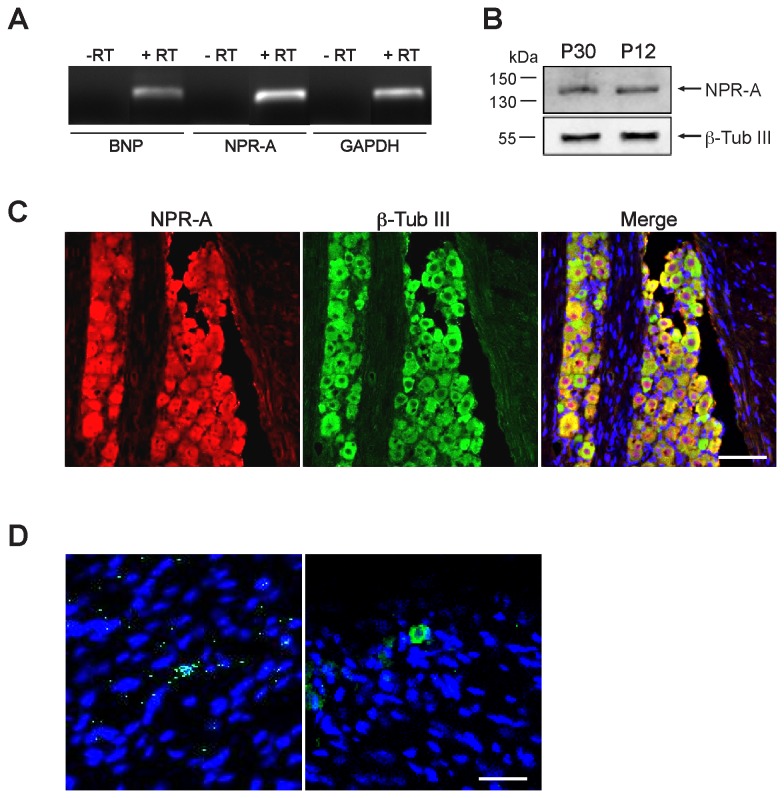
Figure 1. Expression of BNP and NPR-A in adult mouse TG in vivo.
A, PCR products (amplified with 40 cycles) identified by ethidium bromide staining show RT-PCR amplification of BNP and NPR-A mRNA (+RT), as well as of the housekeeping gene GAPDH. Negative control reactions for retrotranscription (-RT) do not show any bands. B, Chemiluminescence image of Western blot showing protein expression of NPR-A in TG from P30 and P12 mice. β-Tubulin III was used as a loading control. C, Example of confocal microscopy images showing the widespread colocalization of immunostaining for NPR-A (red) and β-tubulin III (green). Cell nuclei are visualized with DAPI staining (blue). Scale bar, 100 µm. D, Representative confocal images of longitudinal sections of adult TG. Immunohistochemistry reveals BNP-immunoreactive granules around few nuclei (DAPI). Among these cells, most possess non-neuronal morphology (left panel); neuron-like cells are rarely immunostained (right panel). Scale bar, 30 µm.

